Understanding Brain Scans Before and After EMDR Therapy
Eye Movement Desensitization and Reprocessing (EMDR) therapy has emerged as a powerful tool for treating individuals suffering from trauma and various psychological conditions. This article delves into the intricate relationship between brain activity and EMDR therapy, specifically focusing on the changes observed through brain scans before and after EMDR.
The Science Behind EMDR
EMDR is a psychotherapy treatment developed to alleviate the distress associated with traumatic memories. By utilizing a structured eight-phase approach, this therapy enables patients to process their memories and reshape their reactions. Understanding how EMDR works on a neurological level is crucial for both practitioners and patients.
What Happens During EMDR Therapy?
During EMDR therapy, the therapist guides the patient through focused attention on specific memories while simultaneously engaging in dual stimulation, such as bilateral eye movements. This method is believed to harness the brain's natural processing mechanisms.
Phases of EMDR Therapy
- History Taking: Understanding the patient’s history and identifying targets for reprocessing.
- Preparation: Establishing trust and explaining the EMDR process.
- Assessment: Identifying negative beliefs and developing positive affirmations.
- Desensitization: Utilizing bilateral stimulation to process traumatic memories.
- Installation: Installing positive beliefs related to the trauma.
- Body Scan: Identifying any residual physical tension associated with trauma.
- Closure: Returning to a state of equilibrium and discussing the experience.
- Re-evaluation: Assessing the progress in subsequent sessions.
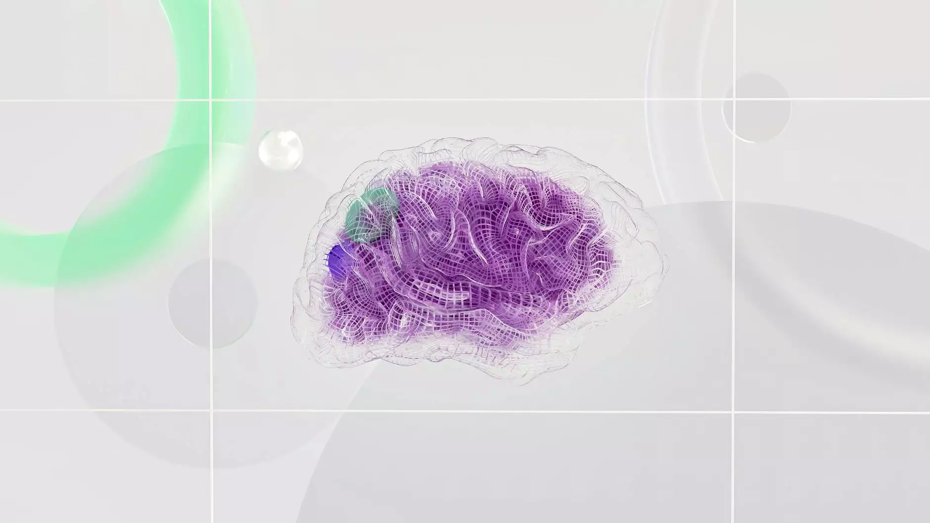
The Role of Brain Scans in Understanding EMDR
One of the most effective ways to observe the impact of EMDR therapy is through functional neuroimaging techniques, such as fMRI (functional Magnetic Resonance Imaging) and EEG (Electroencephalogram). These tools allow researchers to visualize changes in brain activity before and after EMDR sessions.
What Do The Brain Scans Reveal?
Research has shown a significant correlation between EMDR therapy and changes in brain activity, particularly in areas associated with memory, emotion, and cognitive processing. Here are some key findings from studies on brain scans before and after EMDR:
- Reduction in Amygdala Activity: The amygdala, responsible for processing emotions, particularly fear, often shows heightened activity in individuals with PTSD. EMDR has been found to reduce this activity, indicating a decrease in emotional distress.
- Increased Prefrontal Cortex Activation: The prefrontal cortex is associated with higher cognitive functions like decision-making and emotional regulation. After EMDR, increased activity in this area suggests improved emotional processing and control.
- Changes in Default Mode Network (DMN): The DMN is linked to self-referential thoughts and rumination. EMDR appears to alter DMN functioning, which may help individuals detach from negative thought patterns.
Case Studies and Research Insights
A plethora of case studies highlight the effectiveness of EMDR therapy. Various clinical trials have utilized brain scans before and after EMDR to demonstrate its efficacy:
1. Traditional PTSD Cases
In a landmark study conducted by Hofmann et al. (2010), participants with PTSD underwent EMDR therapy while researchers monitored their brain activity through fMRI scans. The results showed a significant activation in the prefrontal cortex post-therapy, alongside reduced amygdala activation, validating EMDR as an effective treatment for PTSD.
2. Chronic Pain and EMDR
Another intriguing study explored the application of EMDR in patients suffering from chronic pain. Researchers observed changes in brain patterns that correlated with pain reduction following therapy sessions. MRI scans indicated changes in neuroplasticity, suggesting that EMDR therapy can promote adaptive changes in brain function.
The Future of EMDR and Neurological Research
The landscape of psychological treatment is continually evolving, and the integration of neuroscience with therapeutic methods like EMDR marks a significant advancement. The ability to visualize how therapeutic interventions affect brain structures is primarily beneficial for practitioners, patients, and researchers alike.
Emerging Trends in Neurotherapy
As neuroimaging technology advances, we expect to see more comprehensive studies on EMDR therapy:
- Personalized Treatment Plans: Utilizing neuroimaging data to create tailored EMDR treatment plans based on individual brain activity patterns.
- Long-Term Effects of EMDR: Investigating the sustainability of brain changes post-therapy and their correlation with long-term recovery.
- Comparative Studies: Comparing EMDR with other therapeutic modalities using neuroimaging to evaluate efficacy.
Conclusion: EMDR as a Catalyst for Change
In conclusion, the use of brain scans before and after EMDR underscores the profound impact of digital therapy on mental health. By bridging the gap between psychological treatment and physiological responses, EMDR not only helps individuals reclaim their lives but also enhances our understanding of the brain’s remarkable capacity for healing and adaptation.
For individuals seeking assistance or looking to explore therapies like EMDR, connecting with qualified professionals is paramount. Indeed, Dr. Eric Meyer, as a skilled psychologist specializing in counseling and mental health, offers invaluable insights and support for those on their journey to recovery. Visit drericmeyer.com to learn more about available services and start your path toward emotional wellness.